|
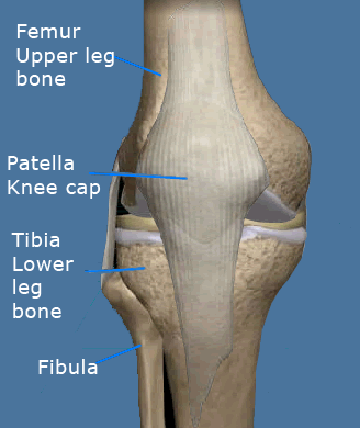
Knee Anatomy
The knee is the largest joint in the body. It is commonly referred to as a "hinge" joint because it allows the leg to flex and extend. While hinges can only bend and straighten, the knee has the ability to rotate and translate.
The knee joint is made up of the femur, tibia, fibula & patella (knee cap). These bones are lined with articular cartilage that allows for smooth motion
of the joint.
Between the tibia and femur lie two fibrocartilages called menisci. The medial (inner) meniscus and the lateral (outer) meniscus rest on the tibial surface cartilage and are mobile. The menisci act as shock absorbers and stabilizers. The anterior cruciate ligament (ACL) and posterior cruciate ligament (PCL) are thick bands of tissue that span the joint from the femur to the tibia.
They guide knee motion and prevent the knee from buckling. The knee joint is surrounded by a capsule that produces synovial fluid to help with smooth motion. Thigh muscles are important secondary knee stabilizers. |
|
 |
| |
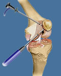
Knee Arthroscopy
Knee arthroscopy is an effective tool for diagnosing and treating knee problems such as meniscus tears, ACL tears, loose bodies and articular cartilage wear. In this procedure, the surgeon examines your knee with an instrument called an arthroscope. An arthroscope is a thin tube with a lens and light on the end that is inserted in your knee through a tiny portal incision. It then projects an image of the inside of your knee onto a TV monitor.
If the suspected problem is confirmed, the surgeon can place small instruments through a second portal, to remove torn or loose cartilage and bone, and repair or reconstruct damaged tendons and ligaments.
An arthroscopy can ultimately provide relief from knee pain and improve mobility. It can be performed as an outpatient under local and/or general anesthesia. Maintaining a normal and active lifestyle with greater comfort is a key benefit of this procedure.
|
|
 |
| |
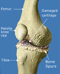
Knee Replacement
In knee replacement surgery, arthritic or damaged bone and cartilage are replaced with metal and plastic surfaces that are shaped to restore knee movement and function. The new artificial knee is called a prosthesis. The prosthesis is generally composed of two metal pieces fitted onto the ends of the tibia (shin bone) and the femur (thigh bone) and a plastic piece inserted between them to act as a bearing. The patella is also resurfaced with a plastic prosthesis.
Stainless steel, cobalt/chrome alloys or titanium may be used for these components. Durable, wear resistant polyethylene (plastic) is used for the bearing and for the patella implant. Bone cement may be used to anchor the prosthesis into the bone. The restoration of knee motion and relief from knee pain that this procedure offers can be life changing. |
|
 |
| |
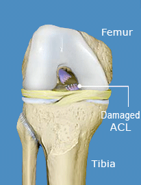
ACL Reconstruction
The Anterior Cruciate Ligament is a stabilizing ligament that connects the femur to the tibia. When torn, the ACL does not heal on its own. Fortunately, reconstructive surgery can help many people recover their full function after an ACL tear, including return to sports.
Surgical treatment usually involves an arthroscopic reconstruction of the injured ligament. The most common type of ACL reconstruction involves harvesting the central third of the patellar tendon to be used as an ACL graft. After performing an arthroscopic examination of the knee, the central third of the patellar tendon is harvested. The remaining tendon is then repaired. Drill guides are then used to place holes in the tibia (bone below the knee) and femur (bone above the knee). When the graft is pulled through the tibial drill hole and into the knee, it will be placed in the same position as the original ACL. After pulling the graft through the drill holes and into the joint, it is then fixed in place with screws. Surgery is followed by early and aggressive physical therapy to restore knee motion and strength.
|
|
 |
|